- Home
- China
- World
- Europe
- Politics
- Business
- Opinions
- Tech & Sci
- Culture
- Sports
- Travel
- Nature
- Picture
- Video
- Live
- TV
- Specials

Share

Copied
"I'm looking for my friend Sam," says Helen Barron, a researcher at the Medical Research Council (MRC) Brain Network Dynamics Unit at the University of Oxford. "Someone tells me that Ben is in the library. I know that Sam and Ben go everywhere together, so I guess that Sam is in the library too."
That is a straightforward example of the kind of 'educated guesses' we make every day to navigate our lives. Until now, we knew very little about how the brain 'joins the dots' between separate memories, but Barron and her colleagues at the University of Oxford have shed some light on the process with their research into brain activity.
The study used both humans and mice, which is unusual but despite the obvious differences, including more complex behavior in humans, the two species are both mammals. According to David Dupret, who also works at the MRC Brain Network Dynamics Unit, humans and mice present a high-level of similarities in their cytoarchitecture, the cellular composition of their central nervous system's tissues.
"We often make decisions where we infer links between different events in our past, but it's not clear how the brain is able to do that," says Barron. "We know that the brain can store memories, but we don't know how it might link together those memories to allow our behavior to be quite flexible."
The research showed that this process takes place in a specific region of the brain, the hippocampus.
"We used MRI in humans, which is a non-invasive method," explains Barron. "And then in mice, we were able to record from lots of cells and also manipulate the activity at the cells by turning the cells on and off using some fancy genetic testing."
In case you missed it:
• The 'superfly' you could soon be eating and COVID-19 hair loss: RAZOR full episode
• How can heat-seeking drones help save wildlife?
• Ground-breaking DNA study busts age-old myths about Vikings
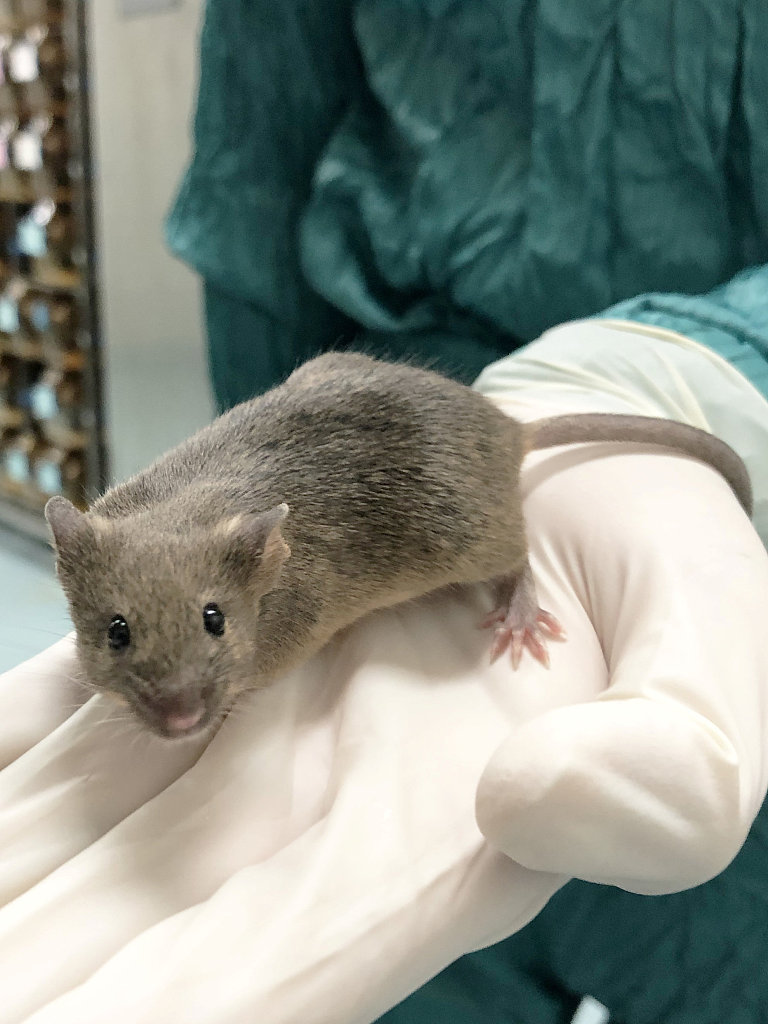
Researchers have confirmed the architecture of a mouse's brain is very similar to humans and has aided studies of the cerebral cortex. /VCG
The people involved in the study were asked to play a game in which hearing a sound signaled that they would also see a colorful picture appear on the wall. They would then play another game in which finding the colorful pictures would be connected to a reward – money.
Similarly, the mice were played a sound before being shown a picture made from LED lights. Then, in a separate exercise, the mice could find a reward of sugar water if the lights were turned on. Both people and mice began connecting the sound with the reward, though there was never a direct link between the two.
But most interestingly, as monitoring on the mice continued through the night, the scientists found that this activity of joining the dots in the brain carried on while the subjects were sleeping.
"What we find is that in periods of rest or sleep, those memories are bound together to form a link that wasn't there before," says Barron. "So we see that two cues that we've never experienced together, a sound cue and a reward, are nevertheless co-activated in sleep. And that represents an inference, a link between two memories that might be useful to the maps in the future."
This might mean that sleeping plays a key role in our creative thinking. "What we're seeing is a new relationship being formed between two memories that have never been experienced together," says Barron.
"That means the brain is generating a relationship that goes beyond what we experience – and I think that's where it relates to creativity, this idea that it's taking you beyond your direct experiences, allowing you to form an insight."
"In England, we are often saying, 'We have to sleep on it'," says Dupret, "like we have an idea and we need to sleep on it. That means that the brain while we're sleeping is actively processing this information, eventually."
The discovery of how the process of putting individual memories together to form new links works in our brain could be useful in treating pathology that affect the memory and specifically the hippocampus.
"The area of the brain we're focusing on, which is called the hippocampus, is the first area to degrade or one of the earliest regions which shows degradation in Alzheimer's disease," explains Barron. "It seems very likely that what we're observing might be a mechanism disrupted in Alzheimer's disease in addition to obvious loss of memory."
Video editor: David Bamford